spinal cord central canal
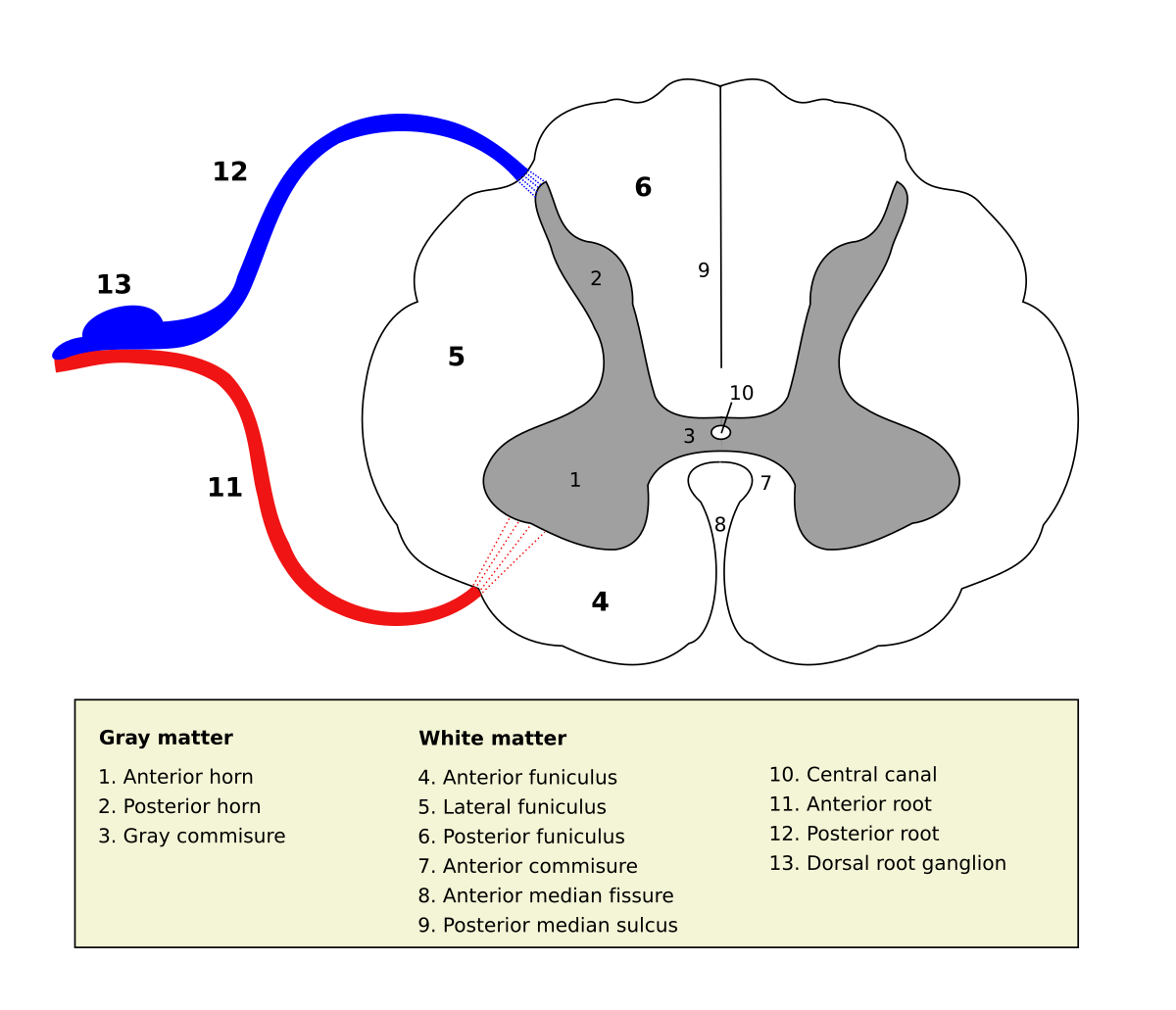 Central canal - Wikipedia
Hydromyelia A hydromyelia should be differentiated from a syringomyelia, which is a cavity filled with fluid inside the spinal cord not lined by ependyma. From: Related Terms: Talia Vertinsky, ... Barton Lane, in , 2008HYDROSYRINGOMYELIAHydromyelia refers to the expansion of the central cord that is lined by ependyma. Syringomyelia refers to an eccentric cystic space that is lined with gliotic tissue. Because hydromyelia and syringomyelia cannot be distinguished by images, the term hydrosyringomielia is often used to cover the two conditions. Patients usually develop hydrosyringomyelia in abnormal CSF flow adjustment, such as Chiari malformation, arachnoiditis, cord injury or chronic compression caused by an intramedular or extramedular mass (Box 10-23). 20,95 In some patients there is no predisponent factor. The hypothesis favored for the formation of sirinx is that the abnormal flow of the FCRs around the fluid of the cordon forces in the cord through abnormal perivascular and extracellular spaces, forming intramedular microcytes that then expand to become cystic cavities. This hypothesis is supported by the so-called pre-syrinx state of the high intramedular T2 signal that can be observed before the formation of the frank cyst. 96 Because hydrosyringomielia can be caused by an underlying neoplasm, the contrast should be administered when the synrinx is first observed (Fig. 10-60). Valerie S. Tay,... Mark Cook, in , 2007SyrinxDefinitionHydromyelia is an abnormal dilation of the central spinal canal, which is usually communicated with the fourth ventricle. A sirinx is a cavity full of fluids inside the spinal cord (syringomyelia) or inside the brain trunk (syringobulbia). A sirinx is described as either communicating or not communicating, depending on whether there is a connection with the CSF tracks. Clinical Characteristics A sirinx may occur in isolation or may be associated with an abnormality in the craniocervical union. A common association is with the type I malformation of Chiari. The level of the sirinx is variable and can in fact be several levels below the increase in the foramen. Syringes can also be secondary for trauma or pathology of the focal spinal cord, including tumors. 25The clinical picture in syringomyelia may be variable. The typical description is of a suspended sensory loss dissociated from committed pain and temperature sensation with intact vibration and sense of joint position, in a capelike distribution on the arms and trunk. The associated motor characteristics include myelopathy, which leads to spasticity and weakness with lower motorized neuron signs at the level of injury and signs of superior neuron engine below the level of the sirinx. A sirinx can manifest with pain, including root pain, neck pain and headaches. Involuntary movements were characterized in 22% of patients with syringomyelia and syringobulbia, including torticollis, mioclonus, tremors, dystony, myoquimy, respiratory sinkinesis and reverse masticatory muscle activity. 26,27Etiology and Pathophysiology The source of fluid in syringomyelia has been postulated to be the same CSF. The exact mechanism for the formation of syringomyelia in a Chiari malformation is not clear. There are two main initial theories that depend on a patent communication between the fourth ventricle and the synrinx: According to the theory "agua-hammer", the CSF pulsatile transmission of the choroid plexo is transmitted through an abnormal fourth ventricle to hammer a dilation inside the spinal cord. Abnormal communication is considered as a result of a delayed and incomplete embryo opening of the fourth ventricle output. According to the theory of "a valve", there are high intermittent pressures generated in the spinal cord, as a result of an unequal pressure gradient during the Valsalva manoeuvre. During the Valsalva manoeuvre, high intratoric pressures are transmitted to the spinal epidural veins, leading to an upward pressure wave pushing CSF from the spinal column to the subarachnoid cranial space. Because there is a downward obstruction to the CSF flow, this pressure is then translated into the CSF movement from the fourth ventricle through the central patent channel to the synrinx. Researchers have re-examined the syringomyelia training mechanism, in which magnetic resonance imaging did not demonstrate a patent communication between the fourth ventricle and the sirinx. Instead, it was postulated that during the systole, the brain expands with blood. This imparts a systolic pressure wave that moves the cerebelous tonsils down and acts on the spine CSF along the surface of the rope to compress the fluid of the rope and the propulsion in the synrinx longitudinally with each pulse. 18,19,25,28ManagementMRI is crucial in the evaluation of a synrinx because it shows the synrinx in itself and associated neurological disorders, including malformations in the craniocervical union (Fig. 38-4). 27 Surgical management consists mainly of drainage or decompression of posterior fossa. Traditionally, surgery involves drainage with the syringostomy and aspiration of the intramedular fluid. Later there was a paradigm shift with magnum foramen decompression, with or without obex plug. More recently, there has been a growing interest in drainage procedures again, mainly due to the advent of micro-surgical techniques and better dazzling materials. Currently, surgery is indicated for a symptomatic sirinx and can be done either with later decompression of fossa or as a shunt procedure. Syringosubarachnoide, syringopleural, or syringopleural position are potentially associated with many complications, including shunt obstruction, dislocation or infection, spinal cord tethering, and septation within the syrinx cavity.27,29Type Jaspan, Rob Dgliineelia, in , 2011Syringomyelia and hydromielia Hydromyelia is characterized by an accumulation of CSF within an expanded central canal of the spinal cord, lined by ependymal cells. Syringomyelia (or syrinx) is the generally used term covering both. In fact, clinical findings and image cannot be differentiated between the two. Syringomyelia may be focal or may be extended to involve the full length of the bone marrow, and is associated with a range of spinal deraffic abnormalities. The ultrasound can enlarge the cord and the normal single central echo (see Fig. 67.25), the previous and later walls of the central channel that are separated by the anecoic fluid. Syringomyelia must be clearly differentiated from a terminalis dilated ventricle, which represents the incomplete embryo regression of the terminal vesicle. The latter occurs immediately cefalad to a normally localized medullaris conus, is not associated with other deraphic anomalies and is not progressive in the follow-up RRM. Alexander de Lahunta DVM, PhD, DACVIM, DACVP, Eric Glass MS, DVM, DACVIM (Neurology), in , 2009 A dilatation of the central channel is hydromielia; a cavitation of the spinal cord parenquima is syringomyelia. Se produce secondary to a herniation of the caudoventral cerebellar vermis through the magnum foramen. The cause of herniation is presumed to be a flowing cranial fose that develops to be too small for the volume of the brain tissue it contains. 5,6,22 to 24 Some veterinarians refer to this as a type 1 Arnold Chiari malformation, which is a human disorder that involves the herniation of the cerebelous tonsils, which are not present in animals. Although pathogenesis can be similar, we believe that this eponym is inappropriate for canine disorder. Until we understand better the cranial malformation that leads to the small cranial fose flow, occipital bone malformation will be considered the primary defect in these dogs. Normally both hydromyelia and syringomyelia are present, and at some point there is a communication between these two spaces filled with liquid. Therefore, we refer to this spinal cord injury as a syringohydromyelia. Syringohydromyelia is most commonly found in the segments of the cervical spinal cord as a multifocal or continuous lesion, but it is also present in the toracolumbar segments. It is believed that it results from the advent of the CSF flow in the magnum foramen caused by the hernia cerebelosa and the compression of the underlying medulla. The cranial cavity is a closed space and the CSF movement is greatly influenced by the pulsation of the arteries associated with the heartbeat. CSF clamps in synchrony with the heartbeat and flows in both directions in the increase of the foramen, but mainly from the cranial cavity to the subarachnoid space of the spinal cord. When this normal pulsating flow is obstructed by the cerebelous hernia, the smooth flow becomes a superior pulsatile flow of pressure and turbulence results in the subarchnoid space of the spinal cord, forcing the CF, the vascular fluid, or both in the parenquima along the penetrating pervascular spaces. The area of less resistance in the spinal cord patch is in the dorsal funiculi, where the synrinx is most commonly found. The pathogenesis of this spinal cord injury in humans, as well as in animals, is very complex and still under intense research. A new concept of physiopathology of the development of syringohydromyelia is based on research that showed a higher pressure of the CSF in the spinal cord cavities than in subarachnoid space. 24 Moreover, these cavities were determined to contain extracellular fluid and not only CSF. The driving force remains the CSF systolic intracranial pressure. This concept is based on the obstruction of CSF flow and the repeated pulsatile mechanical distaste of the spinal cord. The latter results in the accumulation of extracellular fluid due to high pressure in the microcirculation of the spinal cord. Edema in the less resistant dorsal fungi would precede the development of the sirinx. The high-speed CSF jet created by the obstruction of CSF flow paradoxically decreases hydrostatic pressure in subarachnoid space (the Venturi effect). This increases the intramedular distaste of the spinal cord and causes the formation of edema, which leads to syringomyelia. It is well recognized now that the surgical expansion of magnum foramen results in spontaneous resolution of the syringohydromyelia in many patients. Other lesions that cause excessive tissue mass in the brain, especially in the flow cranial pit, such as neoplasms and cysts, also cause the same interference with the CSF flow dynamics and the same spinal cord injuries that resolve after surgical removal of the mass or magno foramen enlargement. Occipital bone malformation is more common in small breeds, especially the Cavalier King Charles spaniel, where there is evidence for a genetic basis. 23 Clinical signs range from a few months to the early adult years and are more commonly related to the cervical spinal cord. 22 The most common clinical sign is discomfort. Affected dogs can be hyperesthetic and act very sensitive to any touch of the head, neck, shoulder or stern. In addition, some dogs exhibit continuous scratches anywhere from their ears to the shoulder region of the chest extremity. This can be related to the tendency of the cervical sirinx to spread laterally from the dorsal funiculus in the adjacent back gray column and interfere with its role in sensory functions. Scoliosis can also result from this spinal gray spinal injury, with the dorsal dorsal spine back neck injury. Some dogs exhibit a lower motor neuron paresis on a chest extremity if the sirinx involves the ventral grey column of the cervical intumeescence on that side. Others, with the participation of the lateral funiculus, can develop a pair of superior neuron and general proprioceptive ataxia. Occasionally, there have been reports of rape signs of ceebello. Facial and sordery paresis has also been reported and seizures rarely occur. Seizures are probably related to pressure alteration in the prosencephalic ventricular system. Once clinical signs are observed, they usually progress until surgery is done to expand the increase in the foramen. The diagnosis is made by the MRI image. The median images of T2 with MR weight are the most successful diagnosis of herniation, as well as syringohydromyelia (Fig. 3-52). Veterinary surgeons have recognized that the thickening of the dorsal dura-arachnoid to the compressed medulla and the first segment of the cervical spinal cord is associated with this chronic hernia. This fibrous tissue is removed in the decompression procedure without apparent clinical complications resulting from surgical manipulation of the cerebel and first segment of the cervical spinal cord or CSF leakage in the adjacent tissues, in our experience. The insertion of a titanium mesh can help protect against the formation of scar tissue on the surgical site. Albert L. Rhoton, Allan J. Hamilton, in , 2005 Alternative Therapies A variety of surgical treatments for hydromyelia associated with Chiari malformation has been proposed (Figure 84.10).21 Gardner10-12 initially recommended a suboccipital craniectomy and cervical laminectomy to decompress malformation and the subsequent oex plug with a small piece of muscle. He subsequently reported that most patients treated in this way developed progressive neurological deficit (personal communication). Plugging the obex increases the possibility of damaging the hypoglossal and vague nuclei that are at the level of the obex near the rhostral end of the central channel. Gardner et al. 13 have also described a procedure called terminal ventriculostomy. The terminal ventricle is the dilated portion of the central channel that extends below the tip of the cone medullaris in the filum terminal (see Figure 84.10). A laminectomy is performed on the flow limit of the fluid sac, and the edge is opened. This procedure does not decompress malformation in magnum foramen, but it may be satisfactory if the patient has symptoms of hydromyelia only. The procedure is inappropriate in cases where the hydromyelic cavity does not extend to the lumbar portion of the spinal cord or to the filum terminal. The CSF takeoff from the side ventricle to the atrium or peritoneum has been considered a treatment mode. 17 However, patients in the first series of the author, even those with marked hydromysylic deficits, did not have significant ventricular dilation. In addition, small ventricles make it difficult to dazzle. It is indicated that rinse is large or if there is greater intracranial pressure. Lumboperitoneal shunting has been defended. It has not been frequently used because of the concern that it may increase the risk of hernia in the increase of the foramen. 19 The percutaneous need of the hydromyelic cavity has been defended as a possible mode of therapy; however, the aspiration of fluid at the time of surgery is followed by the rapid refilling of the hydromyeic cavity of the ventricular system, and it seems unlikely that a needle pathway will remain open.25In 2015Diaglosia directional of the spinal cord It does not usually extend all the way to the cone; it can be sacculated Simulated by truncation artefact or CSF phase ghost drip Metastasis It will normally be detectable in at least 2 Mimicked (or obscured) planes by the CSF pulsation artefact Phase and frequency of the sudden if necessary to confirm as not an artefactAneurysm or Arteriovenous MalformationThe ghost allows the diagnosis of the flow The absence of this artefact in the technically satisfactory MRI test indicates the slow flow or thrombotic injuryCordola bleedingCordogaCaverna malformation, post-raumatic spinal hematoma, mimicked neoplasia by phase ghosts, RF leakage, motion artefact Use recalling echophenium (GRE) to capitalize on the susceptibility artefact In many cases, both hydromyelia and syringomyelia are present (syringohydromyelia). The proximate cause of syringes is likely to alter CSF flow with pressure variations in different parts of subarachnoid space. Pressure variations create forces that lead CSF to the spinal cord. Possible causes include the narrowing of magnum foramen, Chiari I and II malformations, intramedular and extramedular spinal cord tumors, and subarachnoid scarring. The subsequent extension of the cyst may result from rapid changes in intraspinal pressure due to events such as cough or sneezing. Syringohydromyelia's symptoms begin more commonly in late adolescence or early adulthood and progress irregularly, with long periods of stability. The classic presentation is asymmetric and atrophy weakness in the upper limbs, loss of deep reflections of the upper extremity tendon, and loss of pain and sensation of temperature (with preservation of vibration and proprioception) on the neck, arms and upper part of the trunk. With progression, spasticity and hyperreflexia develop in the lower extremities. Progressive ascending and descending levels of sensory weakness and impairment often occur over time. The diagnosis is performed by MHR (see Fig. 426-1). If syringohydromyelia is identified, it is important to perform a brain magnetic resonance to look for associated abnormalities of the craniocervical union. From time to time, the dilation of the mild central channel is discovered incidentally in patients without symptoms or signs of the spinal cord. If no associated cause is found, the prognosis of such anomalies discovered incidentally is generally good. Treatment is directed to the cause, if you can identify. Sometimes the syringopleural or syringoperitoneal cut is performed with variable benefit. In , 2015PATHOLOGYGeneral FeaturesEthologyUnknown No universally accepted unifying theory explains patogenesis Associated abnormalities ± hydromielia, cord tethered, diastematomyeliaChiari 2 malformation; much less common than with myelomeningocele Usually, simple meningoceles is considered an isolated anomaly with the normal level of conus termination However, some skin authors report low conus termination and other spinal anomalies with meningocele and ask if meningocele is a really isolated lesionMeningocele is always covered with the skin may be dysplastic or ulcerated Cervical lesions considered within the cervical myelocytocele Gul Moonis, in , 2008SyringohydromyeliaSyringohydromyelia refers to CSF-filled cavities within the spinal cord (Fig. 6.46). The cystic dilation of the central channel is called hydromyelia, while the liquid cavities within the cord patch are called syringomyelia. Since it is often difficult to make this distinction based on the appearance of image, the general term syringohydromyelia (syrinx) is often used to include all cystic cavities that contain CSF within the spinal cord. Hydromyelia occurs more commonly in association with congenital malformations, more commonly Chiari malformations. Other causes include the narrowing of magno foramen secondary to acondroplasia and tumor. Syringomyelia may be caused by intramedular tumors, traumas, ischemia and arachnoiditis or may be idiopathic. In MRI, syringohidromielia appears as a cystic intramedular collection that usually has CSF signal intensity in all pulse sequences (Fig. 6.46). A high concentration of proteins can increase signal intensity in T1 weighted images. Benign syringes are usually fusiforms in form, with proximal and conical distal margins. Tumor syringes are often more rounded or oval in form. Cavities can be multiple and can be incompletely septated, appearing crowded (Fig. 6.46). The glorious changes may be present at the ends of the synrinx. Improved images by gadolinium are essential to looking for any underlying tumor.142 Recommended Publications: We use cookies to help provide and improve our personalized service and content and ads. By continuing to accept . Copyright © 2021 Elsevier B.V. or its licensors or collaborators. Direct Science ® is a registered trademark of Elsevier B.V.ScienceDirect ® is a registered trademark of Elsevier B.V.

Central canal - Wikipedia
Central canal - definition — Neuroscientifically Challenged

1. Introduction to Syringomyelia - Phenomenology and neurological disorders

Spinal cord | anatomy | Britannica

Cord Cystic Cavities: Syringomyelia and Prominent Central Canal - ScienceDirect
Spinal Stenosis: What is It, Symptoms, Causes, Treatment & Surgery
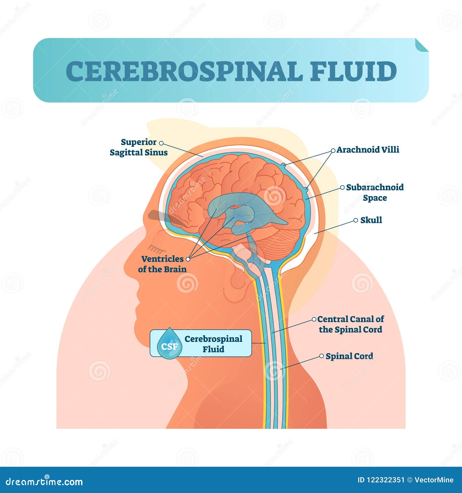
Central Canal Stock Illustrations – 149 Central Canal Stock Illustrations, Vectors & Clipart - Dreamstime
Lab 2 Spinal Cord Histology

Day 172: Know your spinal cord – The lumbar cistern and cerebrospinal fluid | Lunatic Laboratories

Science Source Stock Photos & Video - Spinal Cord, Illustration
2-4. THE SPINAL CORD

Learn About Central Canal | Chegg.com
:background_color(FFFFFF):format(jpeg)/images/library/11512/anatomy-spinal-cord-cross-section_2_english.jpg)
Spinal cord: Anatomy, structure, tracts and function | Kenhub
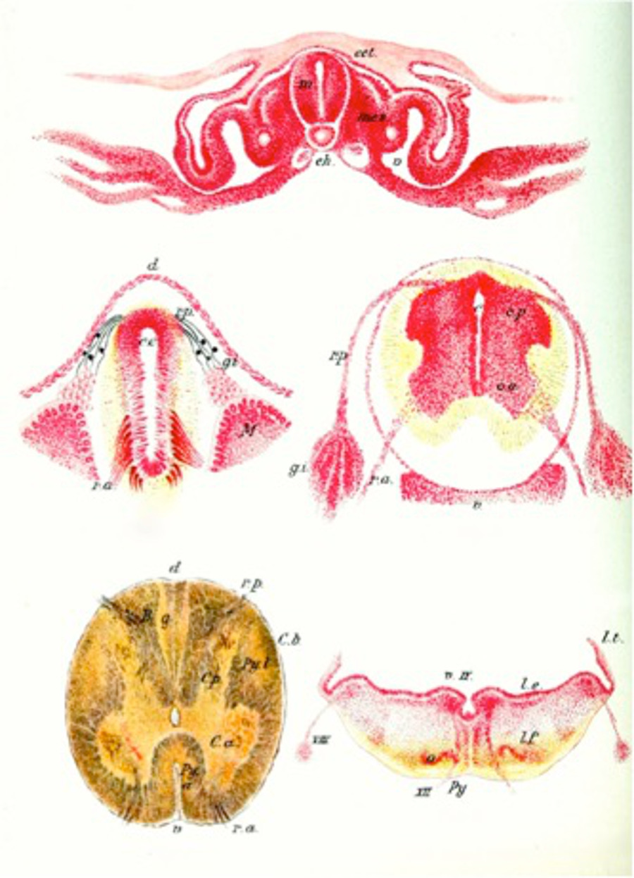
Cureus | The Human Central Canal of the Spinal Cord: A Comprehensive Review of its Anatomy, Embryology, Molecular Development, Variants, and Pathology

Central canal - Wikipedia
Spinal Syrinx Underwriting Case Study

Histological image (H&E) of the human central canal at the center of... | Download Scientific Diagram

Anatomy of Spinal Stenosis
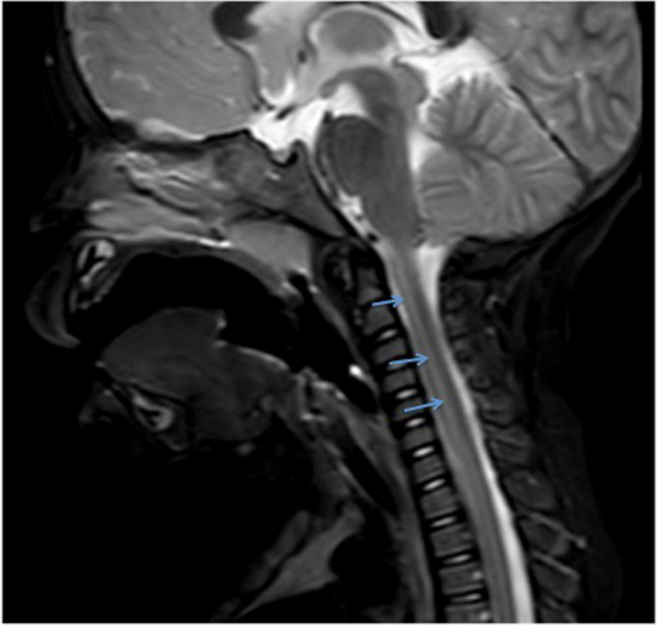
Cureus | The Human Central Canal of the Spinal Cord: A Comprehensive Review of its Anatomy, Embryology, Molecular Development, Variants, and Pathology

Types of spinal stenosis...central canal compresses spinal cord. Lateral and foraminal compress nerve root. | Spinal stenosis, Stenosis, Spinal canal
Cureus | The Human Central Canal of the Spinal Cord: A Comprehensive Review of its Anatomy, Embryology, Molecular Development, Variants, and Pathology
Spinal Stenosis: What is It, Symptoms, Causes, Treatment & Surgery

Pressure-absorption responses to the infusion of fluid into the spinal cord central canal of kaolin-hydrocephalic cats in: Journal of Neurosurgery Volume 58 Issue 2 (1983)

Anatomy | Anatomy of the Spinal Cord and Nerves - YouTube

Ascl1 Balances Neuronal versus Ependymal Fate in the Spinal Cord Central Canal - ScienceDirect

Light Micrograph of the Central Canal of the Spinal Cord In Transverse Section
Lab 1 Neurohistology - Spinal Cord & Meninges
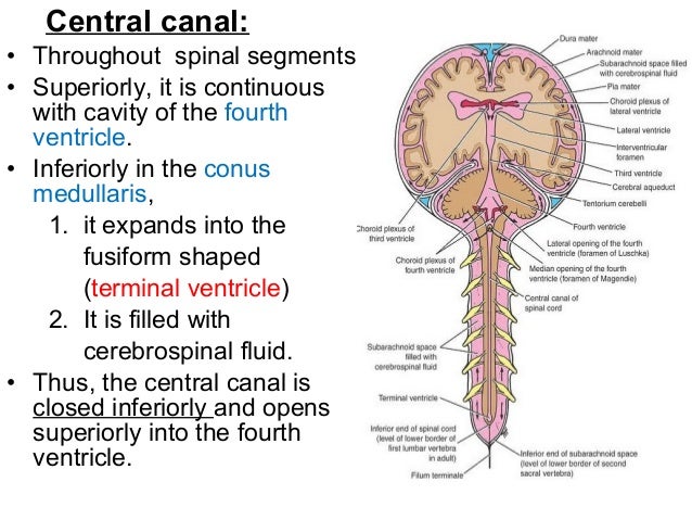
Spinal Cord And Vertebral Canal

The spinal cord central canal: response to experimental hydrocephalus and canal occlusion in: Journal of Neurosurgery Volume 36 Issue 4 (1972)

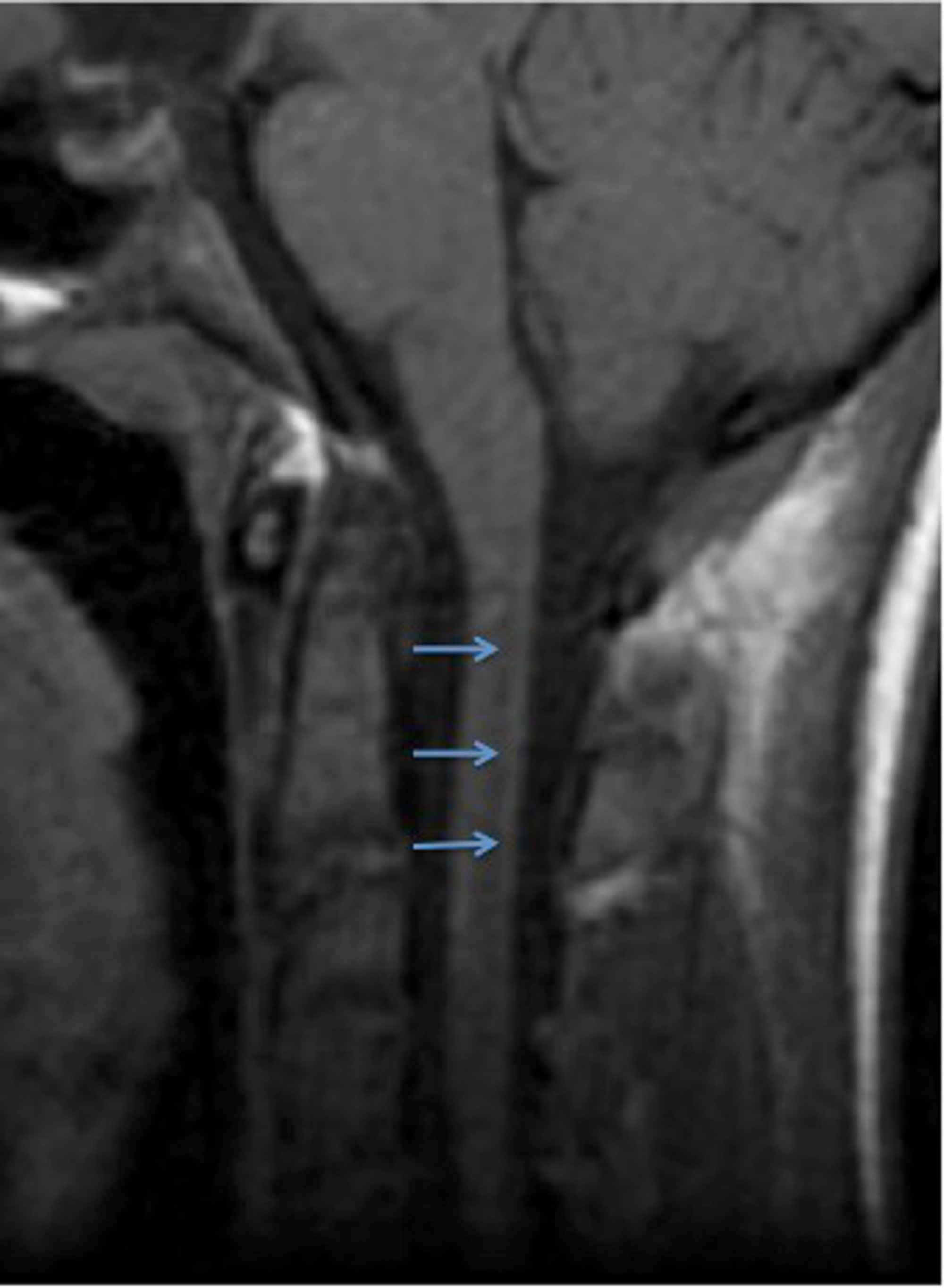
Sagittal MRI scan showing visualization of central canal in the... | Download Scientific Diagram
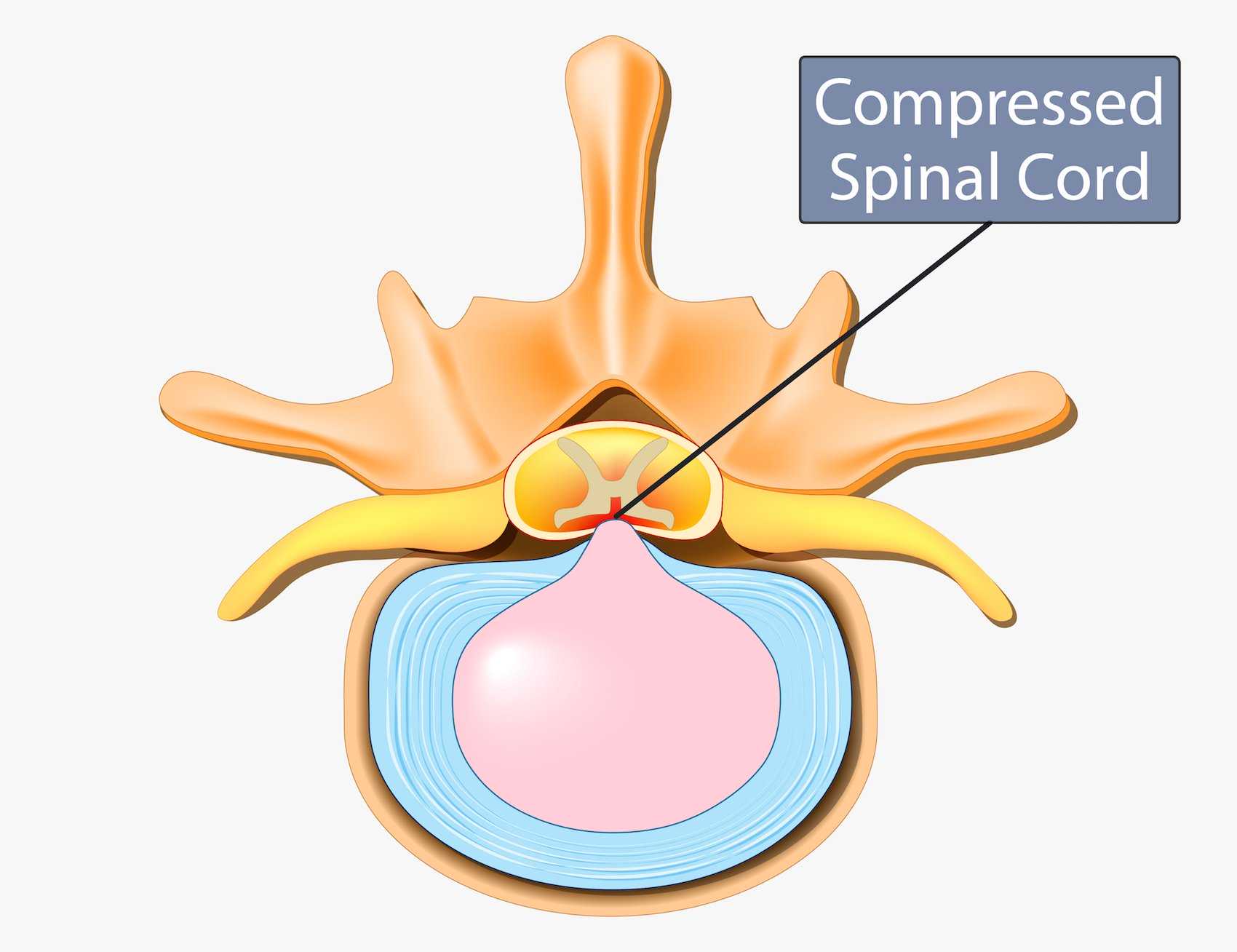
Spinal Stenosis Symptoms, Diagnosis & Treatment | Miami Neuroscience Center
Lab 2 Spinal Cord Histology

Nestin positive cells at the central canal of rat spinal cord... | Download Scientific Diagram

Anterior fissure, central canal, posterior septum and more: New insights into the cervical spinal cord gray and white matter regional organization using T1 mapping at 7T - ScienceDirect

Spinal Cord Gray Matter Anatomy
Spinal Cord

Anatomical continuity of the central canal from medulla through spinal... | Download Scientific Diagram

CH 12 Gross Anatomy of the Spinal Cord
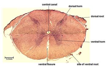
Central Nervous System - Histology - WikiVet English
Cureus | The Human Central Canal of the Spinal Cord: A Comprehensive Review of its Anatomy, Embryology, Molecular Development, Variants, and Pathology
Posting Komentar untuk "spinal cord central canal"